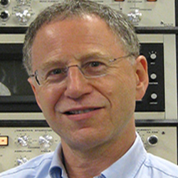
MSE Colloquium - Richard Leapman

When:
Tuesday, March 28, 2017
4:00 PM - 5:00 PM CT
Where: Technological Institute, L361, 2145 Sheridan Road, Evanston, IL 60208 map it
Audience: Faculty/Staff - Student - Post Docs/Docs - Graduate Students
Contact:
Department Office
(847) 491-3537
matsci@northwestern.edu
Group: Department of Materials Science and Engineering (MatSci)
Category: Lectures & Meetings
Description:
Imaging tissues, cells and nanoparticles in 3-D with electron probes
By: Richard Leapman
Abstract: Scanned electron probes can be used to characterize inorganic, organic and biological structures, as well as the interfaces between them in 3-D at the nanoscale. Here, we illustrate the application of scanning transmission electron microscopy (STEM), electron energy loss spectroscopy (EELS), electron tomography (ET), and serial block face scanning electron microscopy (SBF-SEM) to study tissues, cells, and bionanoparticles. The use of focused probes offers important advantages for determining 3-D cellular architecture since it is possible to optimise signals generated by the interaction between the incoming electrons and the specimen. In particular, axial bright-field STEM-tomography, and serial block face SEM are proving valuable for quantitative 3-D ultrastructural analysis of cells and tissues. For example, we have applied these approaches quantitatively to study ultrastructural changes that occur in blood platelets when they are activated, the arrangement of organelles in homone-secreting cells in pancreatic islets of Langerhans, and the interface between osteocytes and mineralised matrix in bone formation. The ability to synthesize multicomponent hybrid nanocarriers with controlled architecture and chemical functionality offers great potential for developing in vivo and ex vivo medical diagnostics and therapeutics. For example, we have used a combination of STEM, EELS and ET to characterise plasmonic gold nanovesicles designed for photothermal therapy, optical nanosensors that detect enzymes expressed by cancer cells, and nanocomplexes taken up by stem cells as magnetic labels for MRI imaging.
Bio: Richard Leapman obtained his B.A. and M.A. degrees in Natural Sciences, and his Ph.D. in physics from the University of Cambridge. He trained as a postdoctoral fellow in the Department of Materials at the University of Oxford, and then under the mentorship of Prof. John Silcox in the Department of Applied and Engineering Physics at Cornell University, where he contributed to the development of electron spectroscopy for the nanoscale characterization of materials. Dr. Leapman subsequently moved to NIH to develop methods based on scanning transmission electron microscopy and electron spectroscopy to determine the structure and chemical composition of cells and supramolecular assemblies. More recently, his group has developed techniques based on STEM tomography for determining 3D ultrastructure in thick sections of cells, as well as serial block face SEM approaches for determining nanoscale tissue architecture. Dr. Leapman received the Burton Medal from the Microscopy Society of America, the Samuel Wesley Stratton Award from the National Institute of Standards and Technology, and the Presidential Science Award from the Microbeam Analysis Society.